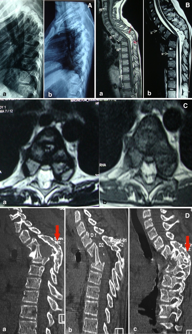
Fig. 1.
a Pre-operative lateral X-ray of dorsal spine (a) show healed TB of spine (D2–4) showing severe kyphotic deformity. Similar post operative X-ray (b) after surgery shows adequate decompression and minimal correction of kyphosis. b T1WI (a) and T2WI (b) midsagittal MRI shows healed vertebral body (D2–4) with severe kyphosis and showing internal salient indenting the spinal cord. The bright signal intensity (T2WI) on spinal cord at apex of kyphosis is suggestive of cord edema. c Axial T1WI (a) and T2WI (b) MRI images show shrunken cord with a cord edema at apex of deformity. d Post-operative reconstructed right parasagittal (a) and left parasagittal (c) image of CT scan show adequate anterior decompression with bone graft in situ. Obliteration of posterior facet joints (solid arrow) suggestive of spontaneous posterior fusion. Similar mid sagittal (b) CT scan image show adequate anterior decompression and interbody bone graft

