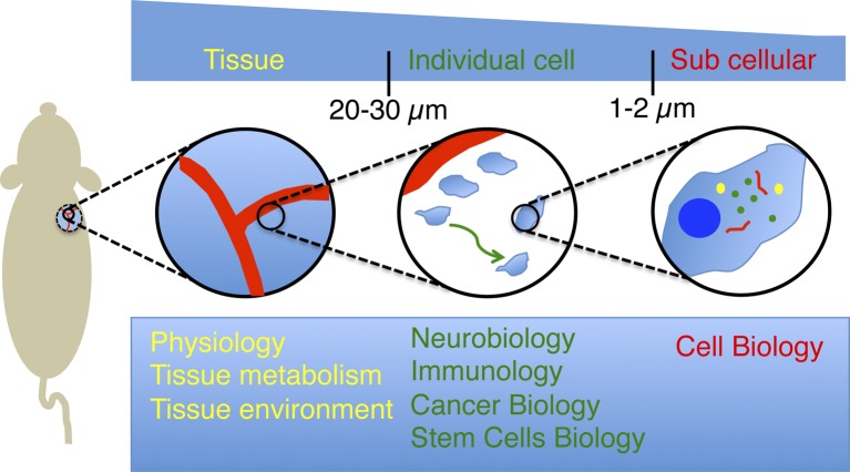
Figure 1.
Spatial resolution and current applications of intravital microscopy. IVM provides the opportunity to image several biological processes in live animals at different levels of resolution. Low-magnification objectives (5–10×) enable visualizing tissues and their components under physiological conditions and measuring their response under pathological conditions. Particularly, the dynamics of the vasculature have been one of topic most extensively studied by IVM. Objectives with higher magnification (20–30×) have enabled imaging the behavior of individual cell over long periods of time. This has led to major breakthroughs in fields such as neurobiology, immunology, cancer biology, and stem cell research. Finally, the recent developments of strategies to minimize the motion artifacts caused by the heartbeat and respiration combined with high power lenses (60–100×) have opened the door to image subcellular structures and to study cell biology in live animals.

