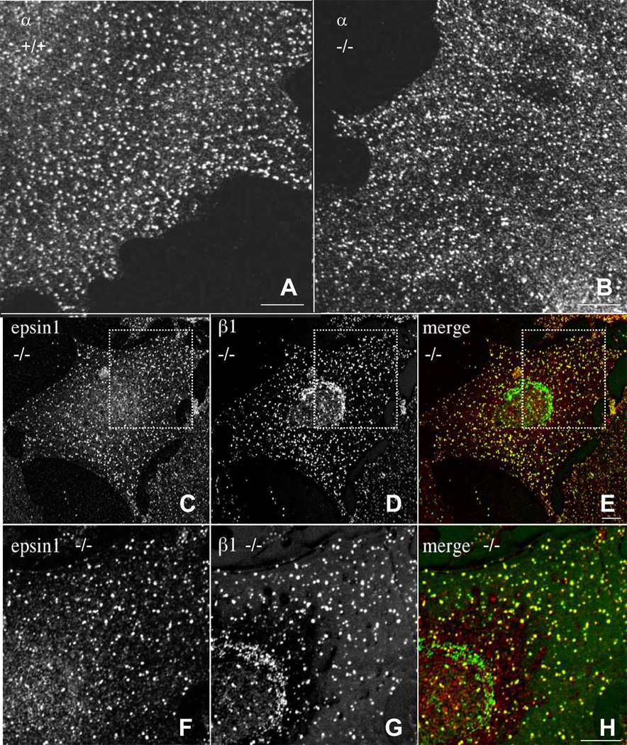
Fig. 6.
β1 subunit is partially incorporated into AP-2 complexes in Ap2β1 mutant mice. (A,B) Wild-type (+/+) and homozygous (Tg/Tg) mouse embryonic fibroblasts were grown on coverslips, fixed in methanol:acetone (1:1), and stained with mouse monoclonal antibodies against α followed by Alexa488-conjugated goat anti mouse IgG. Cells were examined by confocal fluorescence microscopy. Scale bar, 10 µm. (C-E) (Tg/Tg) fibroblasts were fixed and double-stained with antibodies against epsin (red) (C) and β1 (green) (D). Bound antibodies were revealed by Alexa-488 conjugated antibody to mouse IgG and Alexa-555-conjugated antibody to rabbit IgG. All images were obtained by confocal microscopy. Merging images in the red and green channels generated the third panel (E and H) on each row; yellow indicates overlapping localization. Panels F, G, and H are two-fold magnification of the regions shown in panels C, D, and E. Scale bar = 10 µm.

