
Figure 8.
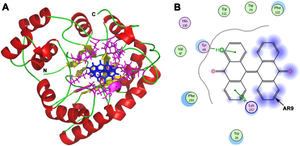
The models of compound AR9 docked into the SjAR structure. A. The predicted overall structure of SjAR bound with compound AR9. The structure of SjAR is shown in a cartoon representation, with α-helices colored red, β-sheets colored yellow and loops colored green. Compound AR9, located in the interior of a hydrophobic pocket, is colored blue. The nearby residues surrounding AR9 are colored magenta and shown with a stick model. View is from the bottom of the (α/β) 8 barrel. B. The interactions between compound AR9 and SjAR. The arrow indicates compound AR9. Polar and non-polar amino acids are colored magenta and green, respectively. Blue background on AR9 structural model indicates the ligand exposure area. The figure is generated with software Molecular Operating Environment 2011 (MOE).