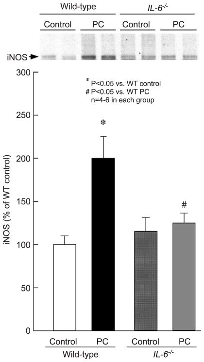
Fig. 7.
Myocardial iNOS expression 24 h after ischemic PC in wild-type and IL-6−/− mice. Myocardial samples were obtained from mice that underwent a sham operation (control) and from the ischemic/reperfused region of preconditioned (PC) wild-type and IL-6−/− mice. Upper panels: Western blot showing that the immunoreactivity of iNOS in the cytosolic fraction increased 24 h after ischemic PC, and that this increase was ablated in the absence of IL-6. Lower panels: Densitometric analysis of immunoreactive iNOS. Data are mean ± S.E.M.

