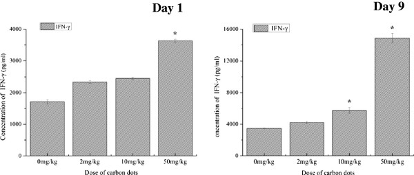
Figure 3.

Influence of carbon dots on splenocyte proliferation of BALB/c mice. BALB/c mice were injected in the caudal vein with different doses of carbon dots. Spleen samples were separated to prepare splenocytes at 1 or 9 days after the administration. T lymphocytes were introduced by ConA, and B lymphocytes were introduced by LPS. Data are presented as means ± standard deviations, n = 5. *P < 0.01 compared with saline group; #P < 0.01 compared with lower dose carbon dot-treated group. Significant difference was calculated by one-way ANOVA using SPSS19.0.
