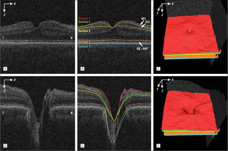
Figure 2.

Segmentation of the nerve fiber layer (NFL), ganglion cell layer (GCL) and combined layer of the outer segment (OS) and retinal pigment epithelium (RPE) from macular and peripapillary spectral-domain optical coherence tomography volumes. A, Flattened and cropped B-scan image of the macular spectral-domain optical coherence tomography volume. B, Image A overlaid with the layer segmentation results. C, Three-dimensional rendering of the surfaces segmented in B. D, Flattened and cropped B-scan image of the peripapillary spectral-domain optical coherence tomography volume. E, Image D overlaid with the layer segmentation results. F, Three-dimensional rendering of the surfaces segmented in image E. N indicates nasal; RPE*, RPE complex; and T, temporal
