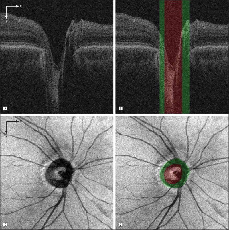
Figure 3.

Segmentation of the optic cup, neuroretinal rim, and neural canal opening from the peripapillary spectral-domain optical coherence tomography volume. A, Flattened and cropped B-scan image. B, Image A overlaid with the cup (green regions) and rim (red region) segmentation results. The neural canal opening segmentation result is the combined region of the cup and rim. C, Spectral-domain optical coherence tomography projection image. D, Image C overlaid with the cup and rim segmentation results.
