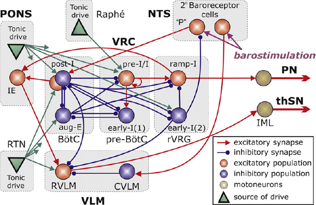
Fig. 3.
The schematic of the extended computational model showing interactions between different populations of respiratory neurones within major brainstem compartments involved in the control of breathing and sympathetic motor activity pons, RTN, BötC, pre-BötC, rVRG, VLM, Raphé, and NTS). A ‘sphere’ represents each population, which consists of 20–50 single-compartment neurones described in the Hodgkin–Huxley style. In comparison with the previous model (Smith et al., 2007), this model additionally incorporates RVLM and CVLM populations in the VLM, an IE phase-spanning population in the pons, and two populations of barosensitive cells in the NTS. The model includes three sources of tonic excitatory drive located in the pons, RTN and raphé shown as green triangles. These drives, especially those from the pontine and RTN sources project to multiple neural populations in the model (green arrows). However,to simplify the schematic, only the most important connections are shown connected to particular populations. The full structure of connections from each drive source (pons/RTN/raphé) to target neural populations of the model and the corresponding synaptic weights are in Table 1 in the Appendix.

