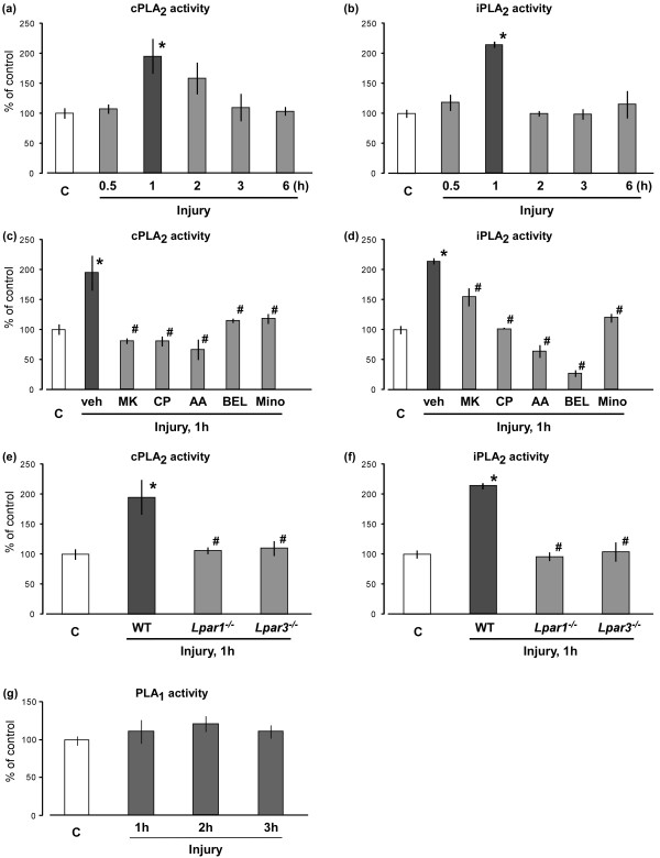
Figure 3.
Blockade of nerve injury-induced cPLA2 and iPLA2 activations. (a and b) Activation of spinal cPLA2 (panel a) and iPLA2 (panel b) were detected by cPLA2 and iPLA2 activity assays at defined time points after nerve injury. The capital letter “C” represents the control group (naive mice). (c and d) After pre-treatments of vehicle, MK-801, CP-99994, AACOCF3, BEL (each 10 nmol, i.t.) and minocycline (30 mg/ml, i.p.) before nerve injury, the activities of spinal cPLA2 (panel c) and iPLA2 (panel d) at 1 h after injury were evaluated. The “veh”, “MK”, “CP”, “AA” and “mino” represent vehicle, MK-801, CP-99994, AACOCF3 and minocycline, respectively. (e and f) Activities of cPLA2(panel e) and iPLA2 (panel f) were measured at 1 h after injury using the Lpar1- and Lpar3-deficient mice. “WT” represents the wide-type mice. (g) PLA1 activity in the spinal dorsal horn was measured by PLA1 activity assay at time-course points after nerve injury. Data represent means ± SEM from experiments using 3-6 mice. *p < 0.05, versus with the control group; #p < 0.05, versus with the vehicle/WT-injury group.

