Figure 6.
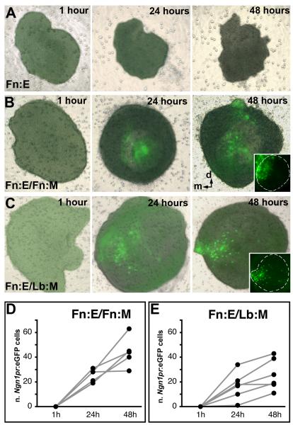
Induction and patterning of Ngn1 in Fn:E by Fn:M and Lb:M. Fb:E is dissected from Ngn1prTg:eGFP E9.0 embryos, and recombined with Fn:M or Lb:M from +/+ E9.0 embryos. Explants have been imaged at 1, 24, and 48 hours after dissection. A) A single Fn:E explant has been imaged at 1, 24 and 48 hours. Ngn1prTg:eGFP is not expressed in Fn:E in the absence of mesenchyme over the 48 hour culture period. B) A single Fn:E/Fn:M explant has been imaged at 1, 24, and 48 hours. Ngn1prTg:eGFP is induced and expressed in Fn:E cells concentrated in the apparent medial aspect of the explant over 48 hours in vitro. The inset at right shows the dense accumulation of Ngn1prTg:eGFP cells in the presumed medial aspect of the explant. C) A single Fn:E/Lb:M explant has been imaged at 1, 24, and 48 hours. Lb:M also induces Ngn1prTg:eGFP expression in cells within the Fn:E. The inset at right shows that the distribution of these cells, however, is more diffuse than in Fn:E/Fn:M explants. D) Numbers of Ngn1pr:eGFP cells increase significantly in Fn:E/Fn:M and Fn:E/Lb:M explants over time (p≤0.001, n=5, Fn:E/Fn:M; p ≤ 0.005, n=5, Fn:E/Lb:M; ANOVA); however, there are significantly more Ngn1pr:eGFP cells in Fn:E/Fn:M explants at 48 hours (p≤0.01;Mann-Whitney).

