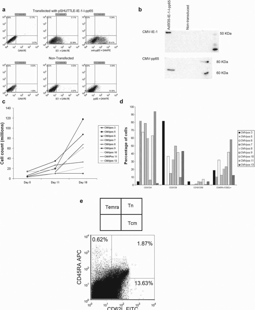
Figure 1.
Expression of CMV-IE-1 and CMV-pp65, T-cell expansion and T-cell phenotype. (a) An intracellular stain of HEK 293T cells transfected with the pSHUTTLE-IE-1-I-pp65 plasmid and stained with anti-IE-1 or anti-pp65. (b) FLY-RD18 cells were transduced with the Ad5f35-IE-1-I-pp65 vector, harvested, and run on a sodium dodecyl sulfate (SDS)–polyacrylamide gel electrophoresis (PAGE) gel. Two identical blots were then probed with anti-IE-1 or anti-pp65 antibodies. (c) Expansion of nine CTL lines to day 18. (d) Reactivity of CTL lines to surface markers for CD3, CD4, CD8, CD16, CD56, CD45RA and CD62L. (e) Terminally differentiated effector memory, central memory and naive T-cell populations after gating on CD3+ and NLVPMVATV (NLV+) cells.

