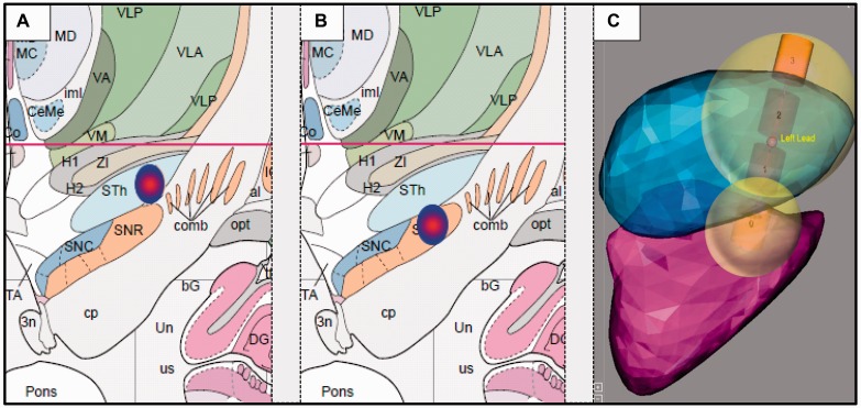
Figure 2.
Localization of active electrode contacts of (A) dorsolateral STN and (B) dorsolateral SNr. Coordinates relative to the midcommisural point (MCP) were: left STN −11.4 ± 0.8, −0.9 ± 2.0, −3.0 ± 1.7; right STN 13.5 ± 1.1, −0.5 ± 1.7, −2.2 ± 1.5; left SNr −10.0 ± 0.9, −3.4 ± 2.1, −6.4 ± 1.8; right SNr 12.1 ± 1.3, −3.3 ± 1.7, −5.8 ± 1.5 (x, y, z; x = medio-lateral, y = anterio-posterior, z = rostro-caudal). Electrode coordinates (mean ± standard deviation in x- and y-direction) are visualized in coronal view on the Atlas of the Human Brain with permission (Mai et al., 2007). (C) An additional illustrative image of electrode localization including a simulation on volume of tissue activated was kindly provided by Medtronic based on work by Yelnik et al. (2007) (atlas) and D’Haese et al. (2012) (atlas and algorithms).

