
Figure 3.
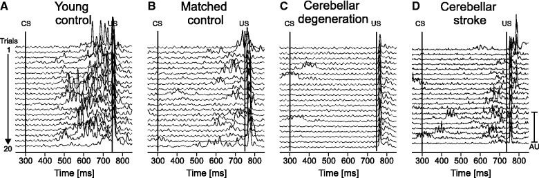
Rectified EMG recordings of orbicularis oculi muscle in (A) a young control (24 years old, male), (B) a matched control (59 years old, male), (C) a patient with cerebellar degeneration (Patient cer-deg-8 in Table 1) and (D) a patient with cerebellar stroke (Patient cer-foc-11 in Table 1). Recordings of the right eye are shown in A–C, of the left eye in D (ipsilateral to the lesion). Each line represents one trial. All 20 trials are shown with the first on top, and the last on bottom of each stack plot. The first line indicates the onset of the conditioned stimulus (CS, ball begins to move) and the second line the onset of the unconditioned stimulus (US, ball touches the forehead).