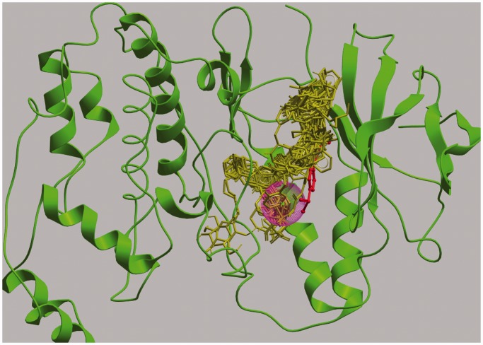
Figure 2.
The binding of ligands in the binding site pocket of various kinases. The Figure shows a superimposition of 81 kinase–ligand complex structures downloaded from the Pocketome server (19), but, for clarity, only one protein structure, MAP kinase p38 (PDB ID: 1a9u), is shown (green ribbons). Ligand molecules are shown as sticks; that for MAP kinase p38 is shown in red and all others in yellow. LISE’s Top1 predicted site (purple sphere) for 1a9u is close to its ligand (red stick), but not close enough to be determined as a successful prediction by the <4 Å distance criterion. This figure was created using the ICM browser (20).

