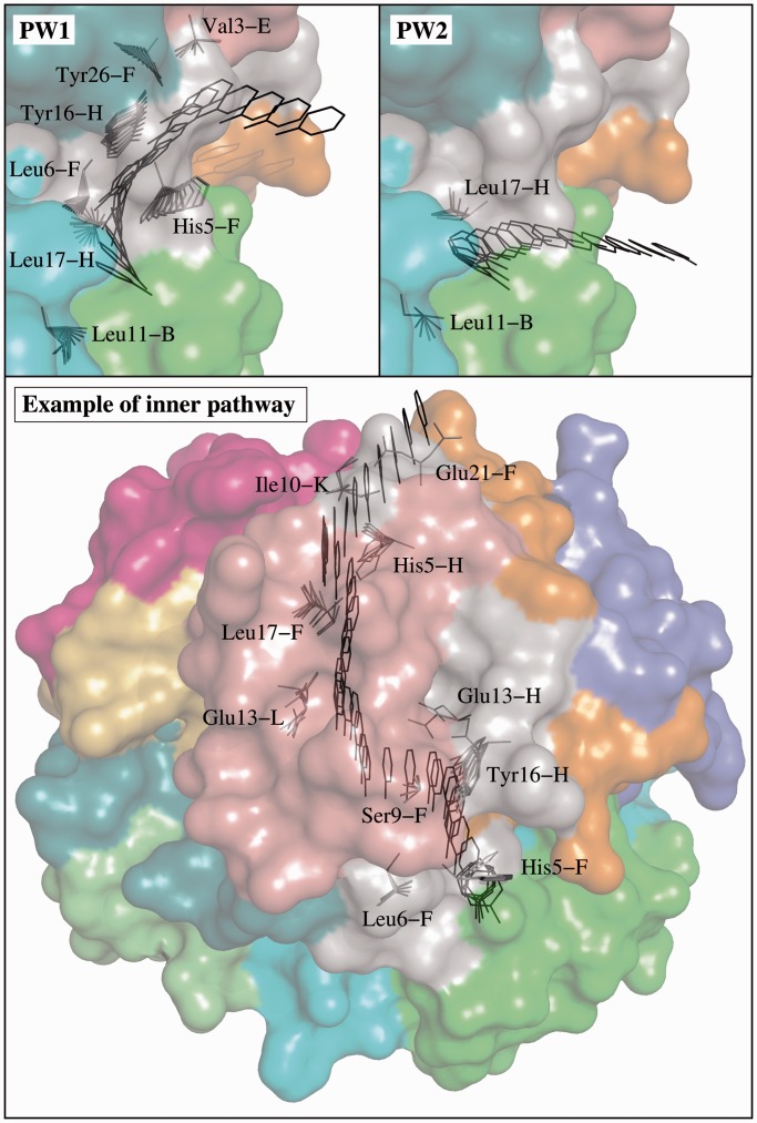
Figure 2.
Different paths for phenol unbinding from the R6 insulin hexamer obtained by MoMA-LigPath. The location of the phenol molecule and the conformations of moving side-chains are represented for some intermediate frames. The two images at the top correspond to paths following the most likely unbinding pathways: PW1 and PW2. The image at the bottom illustrates one of the pathways going through the inner part of the insulin hexamer.

