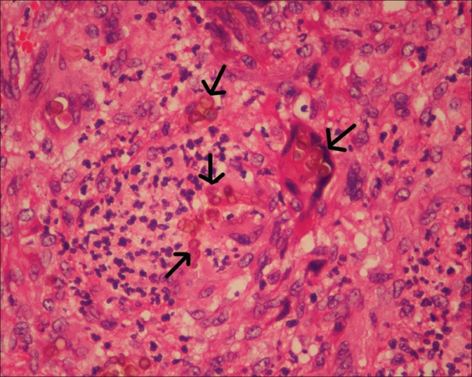
Figure 1.

Light microscopy (× 40, magnification) showing numerous giant cells containing multiple brown-pigmented, ovoid, thick-walled sclerotic/ copper-penny/Medlar bodies seen singly scattered and in chain-like clusters (arrows). These are characteristic of chromoblastomycosis
