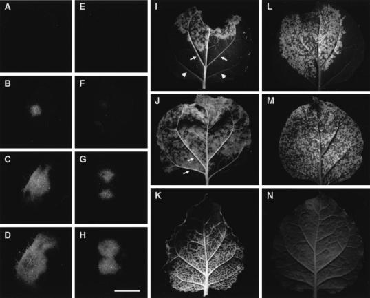
FIG. 5.
Differential local and systemic spread associated with the F12M mutation in TuMV VPg. Virus inocula pCB-TuMV-GFP (A to D and I to K) and pCB-TuMVF12M-GFP (E to H and L to N) were introduced into infiltrated leaf patches with Agrobacterium at a dilution appropriate to give isolated infection foci. The leaves were photographed at 2 (A and E), 3 (B and F), 4 (C and G), 5 (D and H), and 6 (I, J, L, and M) days p.i. Additionally, systemic leaves from infected plants were examined 6 days p.i. (K and N). Passage of the virus in vascular tissues (arrows) is seen for the wt virus only (compare panels I and J with L and M) as fluorescence along the veinal tissues within and beyond the infiltrated area (I and J); compare these with veins from the same leaf (I) without fluorescence (arrowheads). Photographs taken under UV light and printed as greyscale show bright (green) fluorescence against dark tissue background. Bar for panels A to H = 3 mm.

