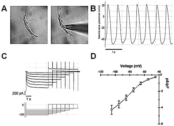
Figure 1.
(A) Dissociated sinoatrial node (SAN) cell in recording chamber before and during patch electrode impalement. Dissociated SAN cells were spindle shaped and more than 80% exhibited spontaneous rhythms in calcium-free enzyme solution. Only single dissociated SAN cells with contractile activity were selected for microelectrode study. (B) Dissociated SAN cells manifest spontaneous action potentials. (C) Representative records from a dissociated SAN cell during the voltage clamp protocol. (D) Summary data: SAN cells exhibit robust If (density 4.3 ± 0.6 pA/pF at −105 mV).

