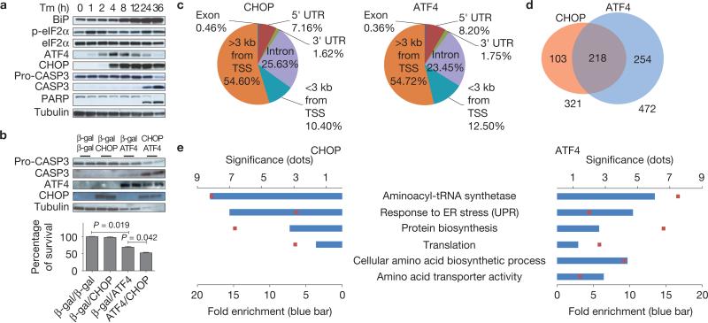
Figure 1.
ATF4 and CHOP bind to promoter regions of genes encoding protein synthesis and UPR functions. (a) Protein expression during ER stress-mediated cell death. Cell lysates were collected at the indicated times after Tm (2 μg ml–1) treatment for western blot analysis. (b) Effect of ATF4 and CHOP expression in WT MEFs. MEFs were infected with adenoviruses expressing CHOP and/or ATF4 at an MOI (mode of infection) of 100. At 24 h after infection, cell lysates were analysed by western blotting (upper panel). At 48 h after infection, cell viability was measured by a WST-8 assay (n = 3 independent experiments; lower panel). (c) Distribution of CHOP and ATF4 ChIP-seq peaks across the genome. The peaks were classified as: within introns (Intron), within 3′ untranslated regions (UTRs), within 5′ UTRs, or within coding sequences (Exon), <3 kb from TSSs or >3 kb from TSSs in intergenic regions. The numbers below the annotations represent the percentage of peaks across the genome. (d) Venn diagram showing overlapping and unique sets of ATF4- and CHOP-occupied genes that have peaks <3 kb from the TSS. (e) Functional enrichment analysis of ATF4 and CHOP target genes that have peaks <3 kb from the TSS. All error bars represent means±s.e.m. Uncropped images of blots are shown in Supplementary Fig. S7.

