Figure 2.

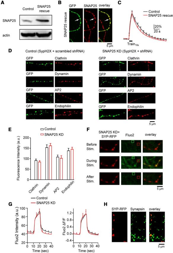
SNAP25 knockdown is specific. A, Western blot of SNAP25 and actin from PC12 cells in control and in cells transfected with SNAP25 shRNA and shRNA-resistant SNAP25 (SNAP25 rescue). B, Immunostaining of SypH2X (antibody against GFP, left, green) and SNAP25 (middle, red) at neuronal branches with (arrow, green) or without (no green staining) triple-transfection of SypH2X, SNAP25 shRNA, and shNRA-resistant SNAP25 (SNAP25 rescue). Right, Left and middle panels superimposed. C, The SypH2X signal induced by Train10s at boutons transfected with only SypH2X (black, control, n = 6 experiments) or with SypH2X, SNAP25 shRNA, and shNRA-resistant SNAP25 (SNAP25 rescue, red, n = 8 experiments). D, Samples of immunostaining against GFP (green, recognizing SypH2X, first and third columns), clathrin, dynamin, AP2, and endophilin (red, second and fourth columns) at boutons transfected with scrambled shRNA and SypH2X (first and second columns, control) or with SNAP25 shRNA and SypH2X (third and fourth columns, SNAP25 KD). E, The immunostaining intensity of clathrin, dynamin, AP2, and endophilin at boutons transfected with scrambled shRNA and SypH2X (control, black) or with SNAP25 shRNA and SypH2X (SNAP25 KD, red). Each group was taken from 2 transfections (clathrin: control, n = 126 boutons; KD, n = 97 boutons; dynamin: control, n = 97 boutons; KD, n = 108 boutons; AP2: control, n = 84 boutons; KD, n = 86 boutons; endophilin: control, n = 91 boutons; KD, n = 95 boutons). Control and knockdown were not statistically significant for each protein. F, Sampled images showing fluo2 signal (calcium indicator, middle column, green) in neuronal branches with or without transfection of SNAP25 shRNA and synaptophysin-RFP (Syp-RFP, left column) at 10 s before (top), during (middle), and 20 s after (bottom) Train10s. Transfection was recognized as red (Syp-RFP) branches. Red and green squares show examples of stimulation-induced fluo2 puncta with and without transfection, respectively. Right, Left and middle columns superimposed. G, Fluo2 fluorescence raw data (left, a.u.) and fractional changes (right, ΔF/F, fluorescence change divided by the mean of baseline fluorescence) induced by Train10s in control boutons (n = 6 experiments, black) and in boutons transfected with SNAP25 shRNA and Syp-RFP (n = 6 experiments, red). H, Antibody staining against RFP (recognizing Syp-RFP, left) and synapsin (middle, bouton marker, green) in a culture transfected with SNAP25 shRNA and Syp-RFP. Right, Left and middle panels superimposed.
