Fig. 2.
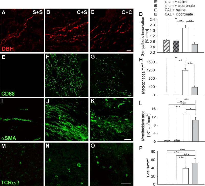
Innervation and inflammatory cell composition of uninjured and injured hearts. Immunostaining of ventricular tissue sections for dopamine β-hydroxylase (DBH) as a sympathetic nerve marker (a–c), CD68 as a macrophage marker (e–g), α-Smooth muscle actin (αSMA) as a myofibroblast marker (i–k), and TCRα/β as a T cell marker (m–o). Sections were obtained from rats receiving sham ligation and saline injections (S+S, a, e, i, m), coronary artery ligation and saline injections (C+S, b, f, j, n), or coronary artery ligations plus clodronate liposomes (C+C, c, g, k, o). Scale bar 10 μm in c and 50 μm in g, k, and o. d Quantitative analysis of sympathetic innervation density as determined by the percentage of section sample area occupied by DBH-ir nerves. h Quantitative analysis of tissue macrophages expressed as the number of CD68-ir cells per mm2. l Quantitative analysis of tissue myofibroblasts as determined by the section sample area occupied by α-SMA-ir. p Quantitation of tissue T cells as determined by the number of TcRα/β cells per mm2. * P < 0.05, ** P < 0.01, *** P < 0.001

