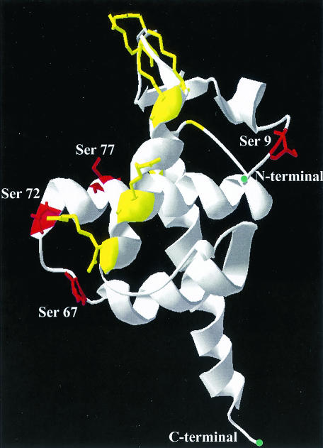
FIG. 10.
Schematic model for MA showing a view of the putative membrane-binding surface as seen from above. Serine residues are shown in red, with their positions depicted, and basic residues, essential for membrane binding, are shown in yellow. The coordinates for this structure were taken from the work of Hill et al. (22), and the model was generated with DeepView/Swiss-PdbViewer software (http://www.expasy.org/spdbv/).

