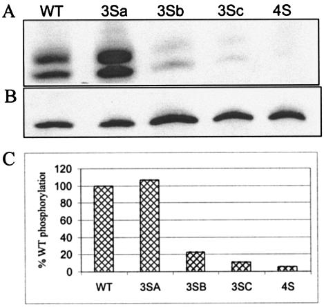
FIG. 9.
Phosphorylation of HIV-1 MA. 293T cells were transfected with WT or mutant proviral clones. Transfected cells were labeled with [32P]phosphoric acid overnight in the presence of Ser-Thr phosphatase inhibitors. The labeled culture supernatants were spun through a 20% sucrose cushion to pellet virus particles. The samples were lysed in RIPA buffer, immunoprecipitated with anti-MA antibodies, separated by SDS-17.5% PAGE, and electroblotted onto a polyvinylidene difluoride membrane. Phosphorylation was analyzed by autoradiography (A and C), and the amount of MA present in each sample was determined by Western blotting with an anti-MA antibody (B).

