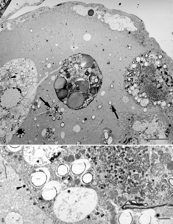
FIG.3.
(A) Low-power electron micrograph of an N2 tick midgut after exposure to a blood meal containing 108 TCID50 of Pr4/ml at 3 weeks postfeeding. Two ASFV-infected PDC with virus factories (arrowheads) are present in the midgut lumen. Free granules (large arrows) are remnants of disrupted PDC. (B) A high-magnification view of the infected PDC attached to the midgut wall with an extensive virus factory (VF) and mature virus particles (arrowhead). Bars, 10 (A) and 1 (B) μm.

