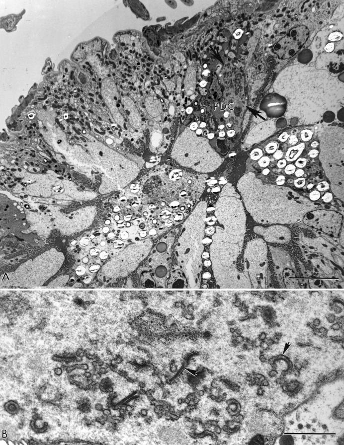
FIG.4.
(A) Low-power electron micrograph of an N2 tick after exposure to a blood meal containing 108 TCID50 of Pr4Δ3-C2/ml at 3 weeks postfeeding. A single infected PDC is found among growing and expanding undifferentiated cells. The typical electron-lucent agranular area of a virus factory (arrows) is present. (B) A higher magnification of the factory region showing developing virus forms (arrowheads) but no mature virions. Bars, 10 (A) and 1 (B) μm.

