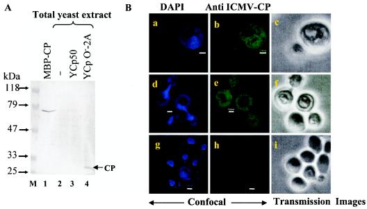
FIG. 4.
Expression of the viral CP in S. cerevisiae transformed with YCpO−-2A. (A) Western blot analysis of IMYMV CP. The total protein was prepared from untransformed and transformed yeast by resuspension of the cell pellet from 1 ml of culture, obtained at the mid-log phase, in 1× sample buffer (0.06 M Tris-HCl [pH 6.8] 10% [vol/vol] glycerol, 2% [wt/vol] SDS, 5% [vol/vol] 2-mercaptoethanol, 0.0025% [wt/vol] bromophenol blue) and boiling for 10 min. The plasmids used for transformation are indicated at the top of each lane (lanes 2 to 4). Fifty micrograms of each protein sample was separated in an SDS-10% polyacrylamide gel and immunoblotted with a heterologous Indian cassava mosaic virus CP antibody. Three hundred nanograms of purified maltose binding protein fusion of CP (MBP-CP) was used as a positive control. (B) Confocal imaging of yeast cells bearing the YCpO−-2A plasmid. The CP was expressed throughout the yeast cells (b and e), and the recognition specificity of the CP antibody was highlighted by the absence of CP staining in the control cells harboring plasmid Ycp50 (h). Panels a, d, and g show nuclei stained with DAPI, and panels c, f, and i show transmission images in white light. Bars, 1 μm. The same sets of cells are shown in rows, and cells at different growth stages are shown in columns.

