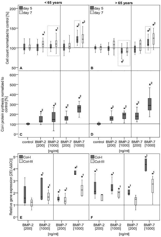
Figure 5. Stimulation potential of TLCs of female donors younger and older than 65 years of age.
A–B Cell count of TLCs of female donors younger (A) and older (B) than 65 years was measured by Alamar Blue assay and given as percentage relative to untreated controls. BMP-2 application increased cell count at day 7 at the low and high concentration in cells of donors younger than 65 years. High BMP-2 concentration significantly decreased cell count at day 5 in TLCs of donors older than 65 years of age. Application of BMP-7 enhanced cell count at the high concentration at day 5 and 7 in cells of both donor groups. Gray boxes in graphs indicate significant differences between donors </>65 years, while older donors showed a decreased stimulated cell count. C–D Col-I protein synthesis in cell culture supernatant of day 7 after growth factor application of TLCs of female donors younger (C) and older (D) than 65 years of age. Col-I synthesis was calculated relative to total protein and given as percentage to the untreated control. The BMP-2 and BMP-7 treatment of TLCs of both donor groups significantly increased Col-I protein synthesis at all concentrations. E–F Col-I and Col-III gene expression after growth factor application of TLCs of female donors younger (E) and older (F) than 65 years. Gene expression is given as 2−ΔΔCt and was normalized to the untreated control. The Col-I and Col-III expression was significantly increased by high BMP-2 and both BMP-7 concentrations in the female group<65 years. In cells of females >65 years both factors increased the Col-I expression, but Col-III expression was only increase by high concentrations of BMP-2 and BMP-7. The asterisks (*) mark significant differences to the untreated control. The numbers give details for the p-value: 1: p≤0.05; 2: p≤0.001.

