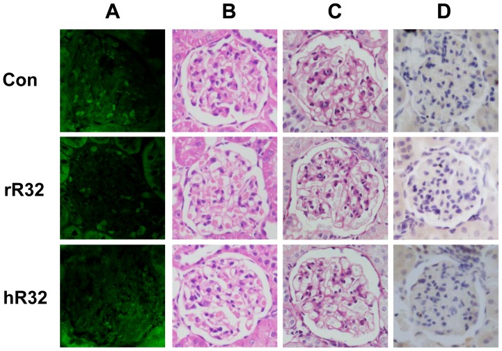
Figure 6. The histological safety assessment in WKYs vaccinated with R32 vaccines.
A–D. The representative kidney images were observed under light microscopy on day 63. A. The IgG deposition in the kidney was not detected in the glomeruli. Both HE (B) and PAS (C) staining showed no obvious pathological changes in the mesangial region of the vaccine groups. D. The immunohistochemical staining against CD14 indicated no macrophages infiltration in the glomeruli. Original magnification: ×400.

