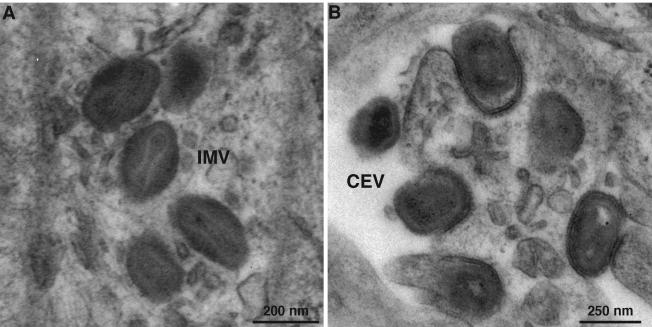
FIG. 8.
Electron microscopy of cells infected with vA28-HAi in the absence of IPTG. BS-C-1 cell monolayers were infected with vA28-HAi in the absence of IPTG for 24 h, fixed, and embedded in EPON, and ultrathin sections were prepared. (A) IMV in the cytoplasm. (B) CEV at the cell surface. The increased electron density on the concave surface of the plasma membrane just under some CEV presumably represents the remains of the fused outer intracellular enveloped virion membrane.

