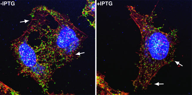
FIG. 9.
Detection of CEV and actin tails by confocal microscopy. HeLa cells were infected with vA28-HAi in the presence (+) or absence (−) of IPTG. After 24 h, the cells were fixed and stained with anti-B5 monoclonal antibody, followed by fluorescein isothiocyanate-conjugated goat anti-rat antibody (green). The cells were then washed and permeabilized prior to staining the DNA with diamidino-2-phenylindole dihydrochloride (blue) and staining filamentous actin with Alexa Fluor 568 phalloidin (red). The arrows point to CEV at the tips of actin tails.

