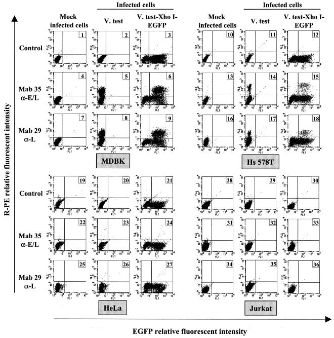
FIG. 1.
Viral gene expression in BoHV-4-infected cells. MDBK (panels 1 to 9), Hs 578T (panels 10 to 18), HeLa (panels 19 to 27) and Jurkat (panels 28 to 36) cells were mock infected or were infected at a multiplicity of infection (MOI) of 0.5 PFU/cell with the BoHV-4 strain V.test or V.test EGFP XhoI. At 30 h postinfection, the cells were harvested and treated for detection of viral gene expression by indirect immunofluorescence staining as described in Materials and Methods. MAb 35 raised against the E-L glycoprotein complex gp6/gp10/gp17 and MAb 29 raised against the L glycoprotein gp11/vp24 were used as the first antibody and were revealed by using PE-GAM. Cells were analyzed by flow cytometry for simultaneous detection of EGFP and PE emissions.

