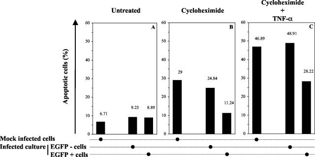
FIG. 2.
Effect of BoHV-4 infection on the susceptibility of HeLa cells to TNF-α-induced apoptosis. HeLa cells were mock infected or were infected with the recombinant strain V.test EGFP XhoI of BoHV-4 at an MOI of 0.5 PFU/cell. At 24 h after infection, apoptosis was induced by CHX-hTNF-α treatment as described in Materials and Methods. The resulting apoptotic effect was assayed 24 h later by using Annexin V-PE labeling, followed by flow cytometry analysis. Panels A, B, and C represent cultures mock treated, treated with CHX, and treated with CHX-hTNF-α, respectively. For infected cultures, the percentage of apoptotic cells was determined for EGFP-positive and -negative cells. Each value represents the percentage of apoptotic cells based on an analysis of 10,000 cells.

