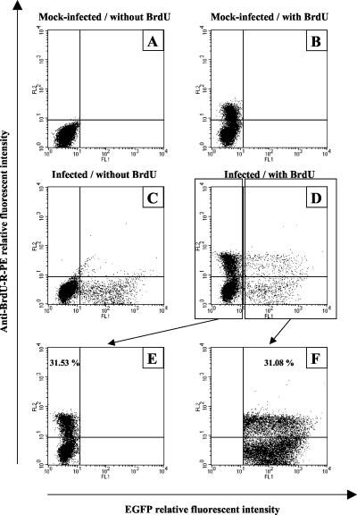
FIG. 4.
Rate of cellular division of EGFP-positive and EGFP-negative cells in HeLa cell culture infected with the BoHV-4 V.test EGFP XhoI strain. HeLa cells were mock infected (A and B) or infected with the BoHV-4 V.test EGFP XhoI strain at an MOI of 0.5 PFU/cell (C and D). The cells were then passaged every other day for 8 days (1:2 split ratio). At 9 days postinfection, the cells were mock pulsed (A and C) or pulsed with BrdU (B and D) for 1 h as described in Materials and Methods. Cells positive for the incorporation of BrdU were then revealed by immunofluorescence staining with anti-BrdU-R-PE and analyzed by flow cytometry for the emission of green (EGFP) and red (anti-BrdU-R-PE) signals. By using the sample of infected cells pulsed with BrdU (D), the rate of BrdU-positive cells was estimated for 10,000 EGFP-negative (E) or EGFP-positive (F) cells.

