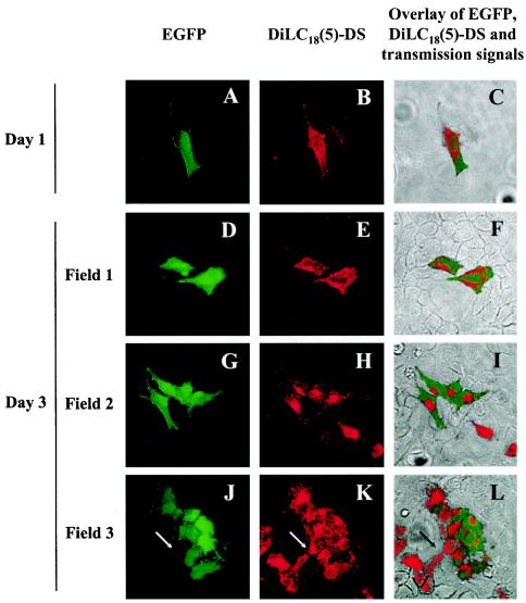
FIG. 5.
Persistence of EGFP expression in the progeny cells of HeLa cells infected with the BoHV-4 V.test EGFP XhoI strain. HeLa cells were infected with the BoHV-4 V.test EGFP XhoI strain at an MOI of 0.5 PFU/cell and then passaged every other day for 6 days (1:2 split ratio). At 6 days postinfection, infected cells were harvested and treated for long-term membrane labeling with DilC18(5)-DS as described in Materials and Methods. Labeled infected cells were then mixed (1:100 ratio) with mock-infected unlabeled cells and grown on glass coverslips. At the indicated times after DilC18(5)-DS labeling, the cells were examined by confocal microscopy for EGFP (A, D, G, and J) and DilC18(5)-DS (B, E, H, and K) signals. Panels C, F, I, and L show the merged EGFP, DilC18(5)-DS, and transmission signals. The side of each panel corresponds to 250 μm of the specimen. The arrows in panels J, K, and L identify the very same point of the examined field.

