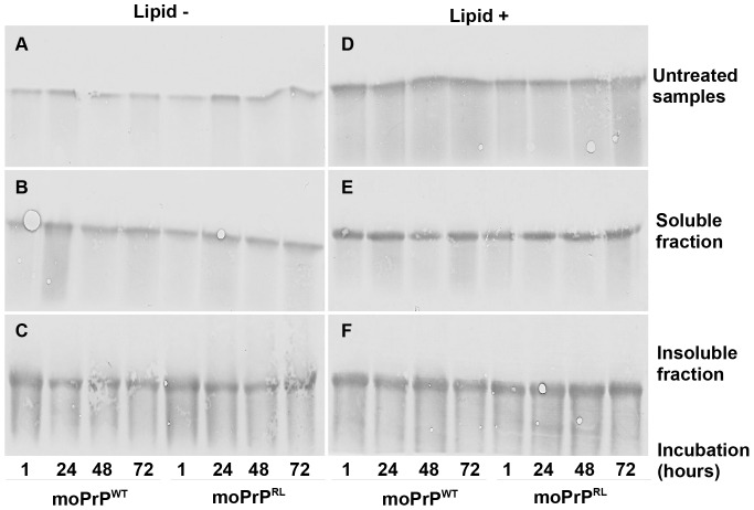
Figure 2. Altered lipid induced misfolding does not result from differences in aggregation between moPrPWT and moPrPRL.
Lipid- (left panels, A–C) and lipid+ reactions (right panels, D–F) containing 100 µg/mL of moPrPWT or moPrPRL were assembled such that all incubations were completed simultaneously. Untreated samples (A and D) were removed and the remaining sample was subjected to centrifugation to separate aggregated (C and F) from soluble PrP (B and E). Samples were then analyzed by Western blot analysis using mAb 4H11. Lipid- samples showed lower intensity than lipid+ samples despite equal starting concentration of protein reflecting that POPG lipid vesicles stabilize PrP at 37°C. moPrPWT and moPrPRL both showed equivalent amounts of soluble (B and E) and insoluble PrP (C and F), indicating that differences in lipid induced generation of PK resistant particles are obviously not the result of differing tendencies to aggregate between the two molecules.

