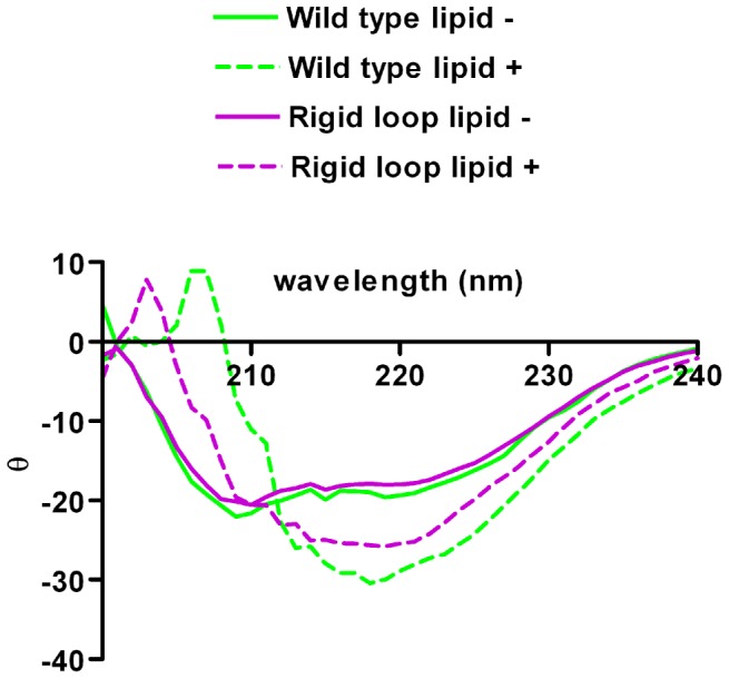
Figure 3. Generation of lipid induced, PK-resistant isoform is accompanied by a structural change increasing β-sheet content.

Differences in secondary structure between moPrPWT and moPrPRL are not detectible by circular dichroism (CD) as shown by their nearly identical CD spectra (solid lines). Upon addition of POPG lipid vesicles, the secondary structure of both moPrPWT and moPrPRL changed to reflect an increase in β-sheet content (dashed lines). In the presence of lipids, moPrPWT and moPrPRL do not exhibit identical CD spectra (dashed lines), possibly indicating differences in lipid induced secondary structural changes between these molecules.
