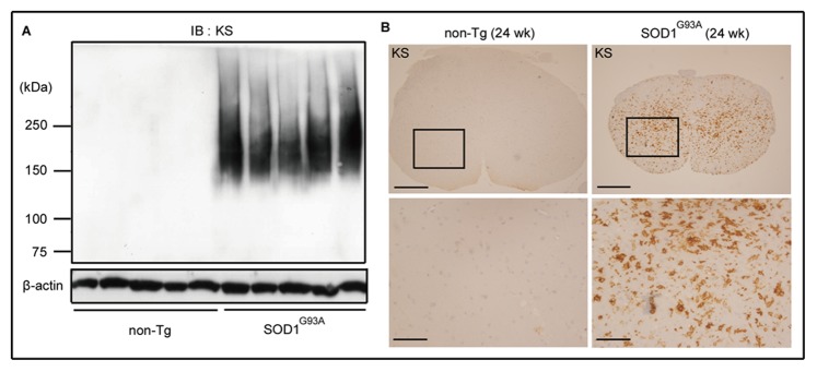
Figure 1. KS expression was induced in the spinal cords of SOD1G93A mice at end stage.

(A) Lumbar spinal cord lysates (24 weeks of age) were subjected to immunoblotting using anti-KS antibody (5D4) (n = 5). β-actin was used as the internal loading control. (B) Lumber spinal cord sections obtained from SOD1G93A mice and their age-matched non-Tg littermates were stained with anti-KS antibody. The lower panels are the highly magnified images of the regions marked with squares. Bars, 200 µm in upper panels; 50 µm in lower panels.
