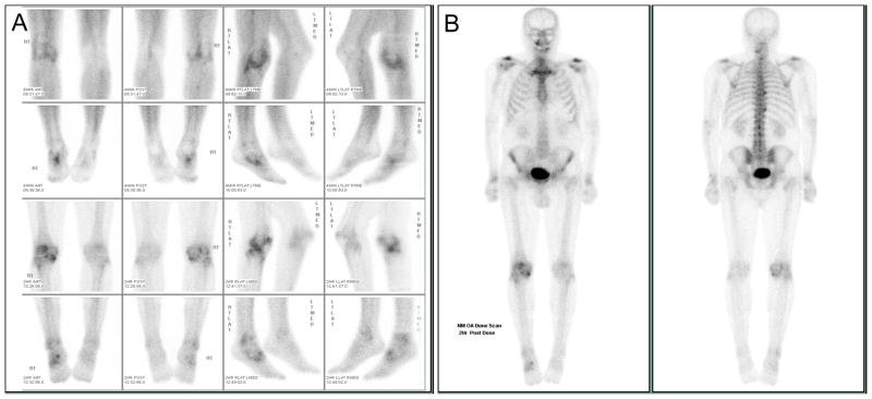
Figure 1. Representative bone scan images of a participant enrolled on the basis of symptomatic radiographic knee osteoarthritis.
A) Spot images of the knees (first and third rows) and ankles (second and fourth rows) obtained starting 4 minutes after injection of the radiopharmaceutical (upper 2 rows) and 2 hours after injection (bottom 2 rows). Abnormal activity is noted in the right knee (medial, lateral and patellofemoral compartments) and in the right ankle and midfoot. B) Whole body bone scan obtained starting 2 hours after injection of the radiopharmaceutical. Abnormal activity is noted in both acromioclavicular, shoulder, and wrist joints, the cervical and thoracic spine, and the right knee, ankle and foot joints as described in A.

