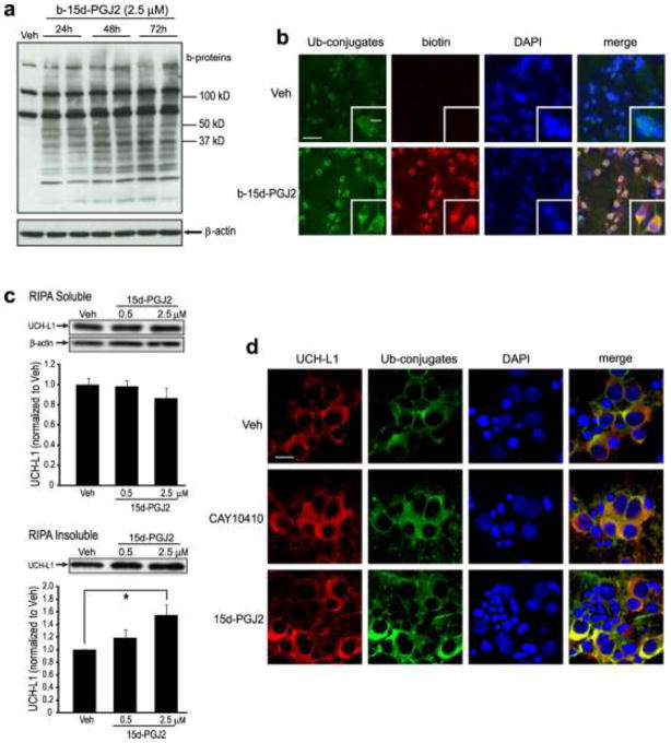
Fig. 6.
Continuous incubation with 15d-PGJ2 modifies endogenous neuronal proteins and increases protein aggregates. (a–b) Rat primary neurons were treated with biotinylated (b-) 15d-PGJ2 or vehicle (Veh). (a) b-15d-PGJ2 adducts to endogenous proteins in primary neurons after treatment for 24 h – 72 h. Cell lysates underwent SDS-PAGE and biotin-incorporated proteins (b-proteins) were detected using HRP-conjugated Streptavidin (upper). β-actin was used as a loading control (lower). (b) Immunocytochemical detection (60X, inset 240X) of ubiquitin and b-15d-PGJ2 using anti-ubiquitin conjugates (Ub-conjugates, green) and anti-biotin (red) antibodies. Blue is DAPI nuclear stain. Scale bar = 60 μm; inset = 20 μm. (c) UCH-L1 aggregates in primary neurons after 15d-PGJ2 treatment. Primary neurons were harvested with RIPA buffer 96 h after treatment with 15d-PGJ2 or vehicle. RIPA -soluble (upper) and -insoluble (lower) fractions were collected, and UCH-L1 was detected by Western blot with anti-UCHL-1 antibody and quantified. Graphs are means +/− SE normalized to vehicle. n=3 per group. * P<0.05 vs vehicle. (d) Immunocytochemical staining (180X) of rat primary neurons 96 h after incubation with vehicle, 2.5 μM CAY 10410 or 2.5 μM 15d-PGJ2. UCHL-1 and ubiquitinated proteins were visualized with anti-UCHL-1 (red) and anti-ubiquitinated conjugates (Ub-conjugates, green) antibodies, respectively. Blue is DAPI nuclear stain. Scale bar = 20 μm. Photos (b,d) were taken with an Olympus confocal microscope

