
Fig. 2.
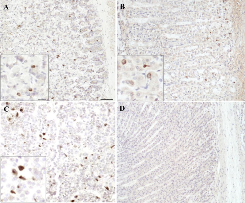
GOAT-immunoreactive cells in the rat and mouse gastric oxyntic mucosa. The anti-GOAT antibody was used for immunohistochemical staining using the DAB method on Bouin’s fixed sections of rat and mouse gastric oxyntic mucosa. The image shows immunohistochemical staining for GOAT in the gastric oxyntic mucosa of mouse in the mid portion (A) and predominantly at the base of the glands in rat (B). GOAT-immunoreactive cells were also observed in the rat pituitary gland (C), whereas no signals were detected in the rat gastric oxyntic mucosa following pre-absorption of the anti-GOAT antibody with GOAT immunogenic peptide (D). The inserts show higher magnifications of the labeled cells. The scale bar in (A) represents 50 μm and 25 μm in the insert.