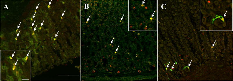
Fig. 3.

Confocal microscopy of doubly labeled oxyntic mucosal sections of mouse and rat. The majority of GOAT-immunoreactive cells (red, A) are located in the mid portion of the mouse oxyntic glands and co-localize with ghrelin (green, arrows, A). In the rat gastric mucosa (B and C), GOAT-immunoreactive cells (red, B) are located at the mid and the base of the oxyntic glands, whereas ghrelin-positive cells (green, B) are mainly seen at the mid portion of the glands. Those cells co-express GOAT and ghrelin (arrows, B). Many of the GOAT-positive cells (red, C) at the base of the glands also express HDC (green, arrows, C). The inserts show higher magnification of double labeled cells. The scale bar in (A) represents 100 μm and 25 μm in the insert.
