
Figure 1.
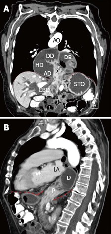
Computed tomographic scan obtained after oral administration of contrast. A: Frontal plane; B: Sagittal plane. The duodenum (D) lies dorsal to the atrial chambers. The descending duodenum is in immediate proximity to the left atrial (LA) chamber. The diaphragm is pointed out as the red dashed line. P: Head of pancreas; CBD: Common bile duct; STO: Stomach; DB: Duodenal bulb; DD: Descending duodenum; HD: Horizontal duodenum; AD: Ascending duodenum; AO: Aorta.