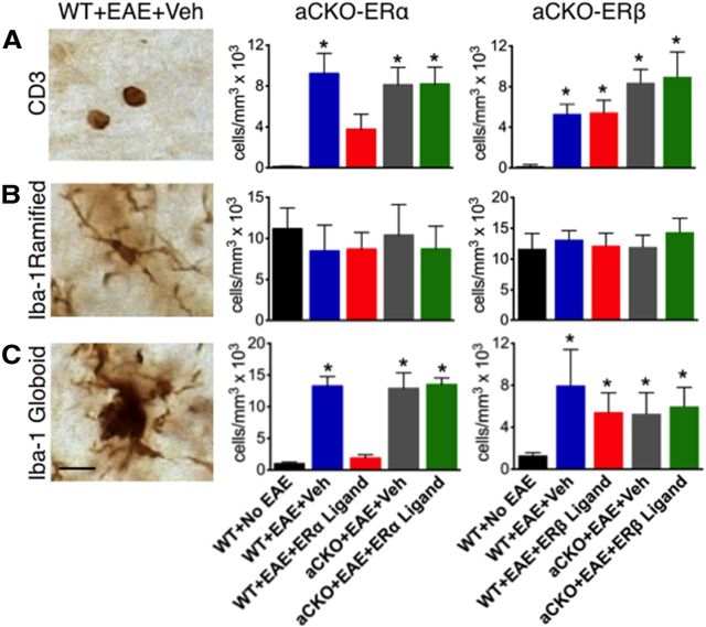
Figure 2.
Quantification of how ERβ, unlike ERα, in astrocytes does not mediate reduction of CD3 T cells and Iba-1 globoid macrophages in EAE spinal cord. A, CD3 T cells were reduced in WT, but not aCKO-ERα, mice with EAE treated with ERα ligand. *p < 0.05 versus WT + No EAE and WT + EAE + ERα ligand (ANOVA with post hoc Bonferroni pairwise analysis). Treatment with ERβ ligand was unable to reduce CD3 T cells in WT or aCKO-ERβ with EAE. *p < 0.05 versus WT + No EAE (ANOVA with post hoc Bonferroni pairwise analysis). B, Iba-1 ramified microglia exhibited no significant difference in number across all experimental groups. C, Iba-1 globoid macrophages were significantly reduced in WT, but not aCKO-ERα, mice treated with EAE treated with ERα ligand. *p < 0.05 versus WT + No EAE and WT + EAE + ERα ligand (ANOVA with post hoc Bonferroni pairwise analysis). Treatment with ERβ ligand was unable to reduce CD3 T cells in WT or aCKO-ERβ mice with EAE. Scale bar, 12 μm. n = 6 per group. *p < 0.05 versus WT + No EAE (ANOVA with post hoc Bonferroni pairwise analysis).

