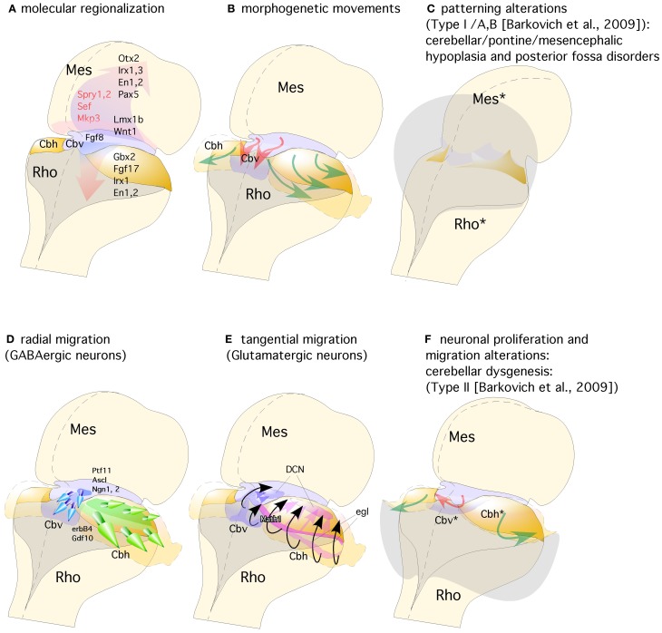
Figure 3.
Representation of a dorsal view of mid-hindbrain junction, where the isthmic segment is colored in light purple (which will generate also cerebellar vermis: Cbv) and the cerebellar plates in yellow (which will generate cerebellar hemispheres). (A) The isthmic organizer expressing Fgf8 (pink arrows) induces the expression of Sprouty, Sef, and Mkp3 in this region; and is also required for other genes differentially expressed in the midbrain (Mes) or rhombencephalic (Rho) neuroepithelium. (B) The spatio-temporal expression of these genes regulates the normal morphogenesis and growth of the cerebellar vermis (Cbv; red arrows) and hemispheres (Cbh; green arrows). (C) Failure in proper isthmic organizer development (due to a lack of morphogenetic signaling or disruption of gene expression) can result in cerebellar (Rho*) and mesencephalic (Mes*) hypoplasia due to a strong increase of cell death with posterior fossa disorders and fourth ventricle dilatation (gray shadow). (D) Radial migration of GABAergic neurons in the cerebellar vermis (Cbv; blue arrows) and hemispheres (Cbh; Green arrows). (E) Rhombic lip specification is regulated by Math1. Tangential migration of glutamatergic neurons of the deep cerebellar nuclei (DCN) and granule cells (egl) are represented by pink and black arrows, respectively. (F) When normal development of cortical cerebellar cells is disrupted, the structural phenotype is classified as cerebellar dysgenesis (Cbv* and Cbh*), with enlargement of the fourth ventricle and reversion of cerebellar-choroidal junction (arrows).

