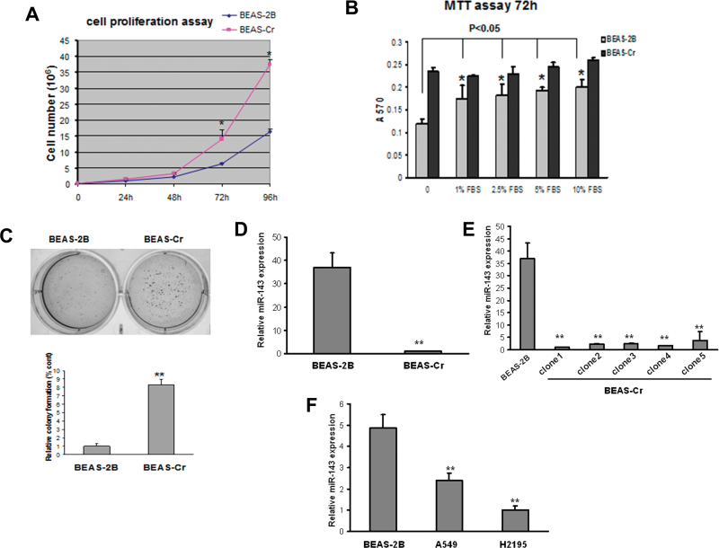
Fig. 1.
miR-143 is downregulated in Cr (VI)–transformed BEAS-2B cells and human lung cancer tissues. (A) An equal number of BEAS-2B and BEAS-Cr cells (10,000 cells) were seeded in 12-well plates to analyze cell proliferation by counting with trypan blue exclusion for every 24h. (B) Cells were seeded in 96-well plates and cultured in medium with different concentrations of serum. 3-(4,5-Dimethythiazol-2-yl)-2,5-diphenyl tetrazolium bromide assay was performed at 72h to assess the cell proliferation rates. (C) An equal cell number of cells (5000) were seeded in a six-well plate and cultured in soft agar and incubated at 37°C for 2 weeks. Representative images for BEAS-2B and BEAS-Cr are presented (left panel). Number of colonies from soft agar assay was counted for each group shown in lower panel. (D–F) miR-143 expression levels were determined by Taqman RT-qPCR in parental Cr (VI)–transformed BEAS-2B cells (BEAS-Cr) and different BEAS-Cr clones. Relative miRNA expression levels were represented as RQ using  methods. The values were normalized to the U6 expression level and that in BEAS-2B cells. Mean ± SE values were from three separate experiments. **Significantly different compared with control (p < 0.01).

