Fig. 3.
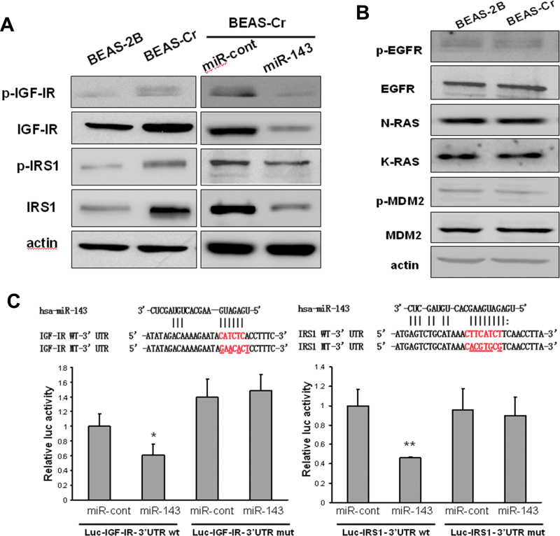
miR-143 targets both IGF-IR and IRS1 in BEAS-2B cells. (A) Total epidermal growth factor receptor, N-Ras, K-Ras, and MDM2 protein levels along with their protein phosphorylation levels were determined by immunoblotting in BEAS-2B cells and BEAS-Cr cells. (B) Basic expression levels of p-IGF-IR, IGF-IR, p-IRS1, and IRS1 were determined by Western blotting in BEAS-Cr cells, BEAS-As cells, and the passage-matched control BEAS-2B cells (left). BEAS-Cr and BEAS-As cells were transiently transfected with 25nM miR-143 or negative miR control precursor. IGF-IR and IRS1 expression levels were determined by Western blotting (right). (C) Top: Sequence alignment of human miR-143 with 3′-UTR of IGF-IR or IRS1. The mutation sites in the 3′-UTR of IGF-IR and IRS1 were shown in the third row for creating the mutant luciferase reporter constructs. The luciferase activities were presented as relative luciferase activities normalized to those of the cells cotransfected with wild-type 3′-UTR reporter and miRNA precursor control. * indicates significant decrease compared with that of control cells (p < 0.05). All tests were performed in triplicate and presented as mean ± SE.

