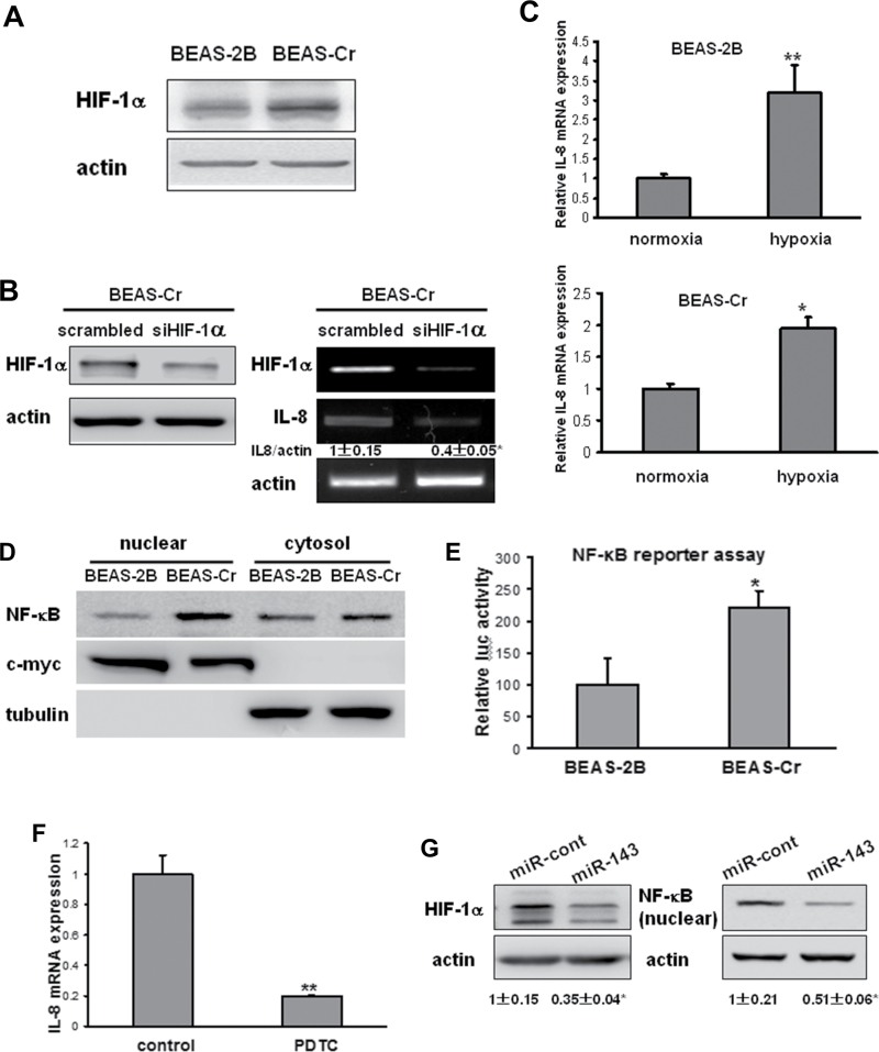
Fig. 7.
Induction of IL-8 expression is mediated by HIF-1α and NF-κB. (A) The HIF-1α protein levels were determined by immunoblotting. (B) BEAS-Cr cells were transfected with 50nM of an siRNA scramble control or SMARTpool siRNAs against HIF-1α for 60h. Left panel: HIF-1 protein expression was assessed by immunoblotting. Right panel: total RNAs were extracted and subjected to RT-PCR analysis for HIF-1α and IL-8 expression levels. (C) BEAS-2B and BEAS-Cr cells were cultured in a hypoxic chamber for 24h. Cells cultured under normoxia condition were served as control cells. RNAs were extracted and subjected to SYBR-Green RT-qPCR for IL-8 mRNA expression levels. (D) NF-κB p65 expression was assessed in nuclear and cytosolic fractions of BEAS-2B and BEAS-Cr cells. The nuclear marker c-myc and the cytosolic marker tubulin were used as loading control. (E) Cells were seeded in 12-well plates and transiently transfected with human NF-κB luciferase reporter and β-galactosidase plasmids. The cells were cultured for 48h after transfection, and relative luciferase activities were measured as described in Materials and Methods section. (F) BEAS-Cr cells were treated with or without NF-κB inhibitor PDTC (40μM) for 24h. Total RNAs were extracted and subjected to qRT-PCR analysis for IL-8 expression levels. The experiments were performed in triplicate and presented as mean ± SE. ** indicates significant difference compared with control group (p < 0.01). (G) BEAS-Cr cells were transiently transfected with miR-143 or miR control precursor for 70h. The expression levels of HIF-1α in total protein extracts and NF-κB levels in nuclear fraction were assessed by immunoblotting.

