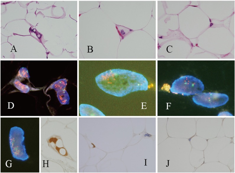
Figure 2.

An ALT/WDLS with typical lipoblasts (case 1, A) and atypical lipocytes (case 2, B), and one tumor that cannot be distinguished from a benign lipoma without the use of FISH (case 3, C). FISH with a probe directed against MDM2 demonstrated that MDM2 is amplified (D-F: orange fluorescence, MDM2; green fluorescence, centromere 12). Dual color FISH with probes directed against MDM2 (orange fluorescence) and CDK4 (green fluorescence) demonstrated that both genes are amplified (G: the 2 nuclei overlap). Analysis of MDM2 expression by IHC revealed unequivocal nuclear staining (H), heterogenous staining (I: positive and negative nuclei), and negative staining (J). (Case 1: A, D, G, and H; Case 2: B, E, and I; Case 3: C, F, and J).
