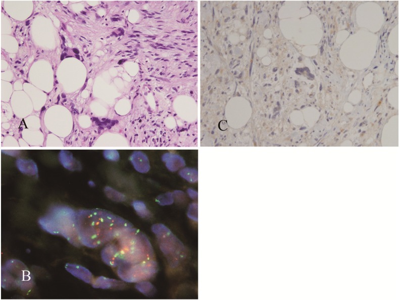
Figure 4.

Pleomorphic lipoma showing marked nuclear atypia (A). FISH analysis with a probe directed against MDM2 (orange fluorescence) and centromere 12 (green fluorescence) detected nuclei with numerous MDM2 signals that were closely associated with centromere 12 signals (B). IHC analysis did not detect nuclear overexpression of MDM2 (C).
